Application of ultrasonography for sex identification and reproductive assessment in Nile tilapia
Distinct echogenic patterns allow for accurate differentiation between female and male fish
Improved breeding programs are important for the global tilapia farming industry, including those for accurate sex determination to inform selective breeding program and population management. Indeed, the optimal male-to-female ratio for effective tilapia reproduction is generally one male to two to four females, as a single male can produce sufficient sperm to fertilize the eggs of several females. Accurate sex selection is, therefore, essential for precise production planning, ensuring that fry output aligns with production goals.
The conventional method currently used for sex selection in tilapia relies on an examination of the fish genitalia, which is heavily dependent on the experience of the workers and can lead to errors. In fact, manual sex sorting based on external genitalia has an accuracy rate ranging from 80 to 90 percent, which results in a high ratio of misidentified fish. Sex differentiation in tilapia can be done at the farm level when the fish are four to six months old and have an average body length of 20–30 cm.
Ultrasonography is a diagnostic imaging technique in which high-frequency sound waves are used to produce real-time images of internal organs, tissues, and blood flow. The technique has been successfully established in various animal species to monitor gonadal development, assess reproductive status, and detect abnormalities of the reproductive organs. Ultrasonography can be used to monitor the spawning status of fish and thus allows for the more efficient management of broodstock. It has been applied for sex identification and to assess the maturity stage of various species of fish, but it has not yet been applied for sex selection in tilapia or to distinguish between female and male tilapia.
This article – summarized from the original publication (Setthawong, P. et.al. 2024. Ultrasonography for non-invasive sex identification and reproductive assessment in Nile tilapia (Oreochromis niloticus). Front. Vet. Sci., 08 October 2024, Sec. Animal Reproduction – Theriogenology, Volume 11 – 2024) – reports on a study that evaluated the application of ultrasonography for sex identification and reproductive assessment in Nile tilapia.

Study setup
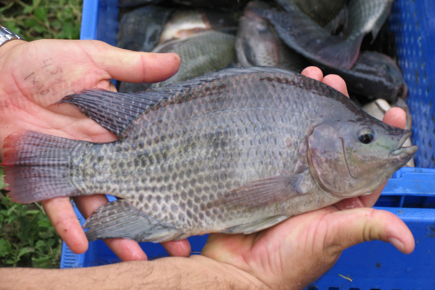
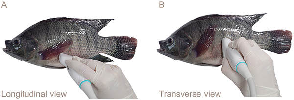
Healthy Nile tilapia (232–1,281 grams) were randomly acquired from a hatchery in Phetchaburi Province, Thailand. Ultrasonography was performed on 23 anesthetized fish to capture longitudinal and transverse images of the ovaries and testes. Female tilapias were identified by the presence of numerous small egg granules and gray or light gray ovarian tissue and male tilapia by the homogeneous echogenicity and uniform gray tubular appearance of the testes. These distinct echogenic patterns allowed for accurate differentiation between the female and male fish.
For detailed information on the experimental design, fish husbandry and handling, ultrasonography data collection and interpretation, refer to the original publication.
Results and discussion
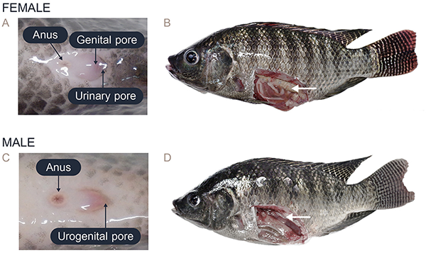
In this study, the female Nile tilapia had a mean weight of 511.63 ± 278.90 grams and a body length of 29.56 ± 4.4 cm, while the male tilapia had a mean weight of 726.14 ± 371.78 grams and a body length of 32.57 ± 4.35 cm. Sex identification was determined by analyzing the external morphological characteristics of the fish. The females were distinguished by their oval-shaped external genitalia, which featured three distinct openings: the anus, genital pore, and urinary pore (Fig. 2A). During spawning, the females showed abdominal distension and enlargement (Fig. 2B). In contrast, the external genitalia of the male fish had a small cone shape with two openings: the anus and urogenital pore (Fig. 2C). The testes were located dorsally to the digestive tract within the coelomic cavity, and the appearance and size varied depending on the reproductive stage (Fig. 2D).
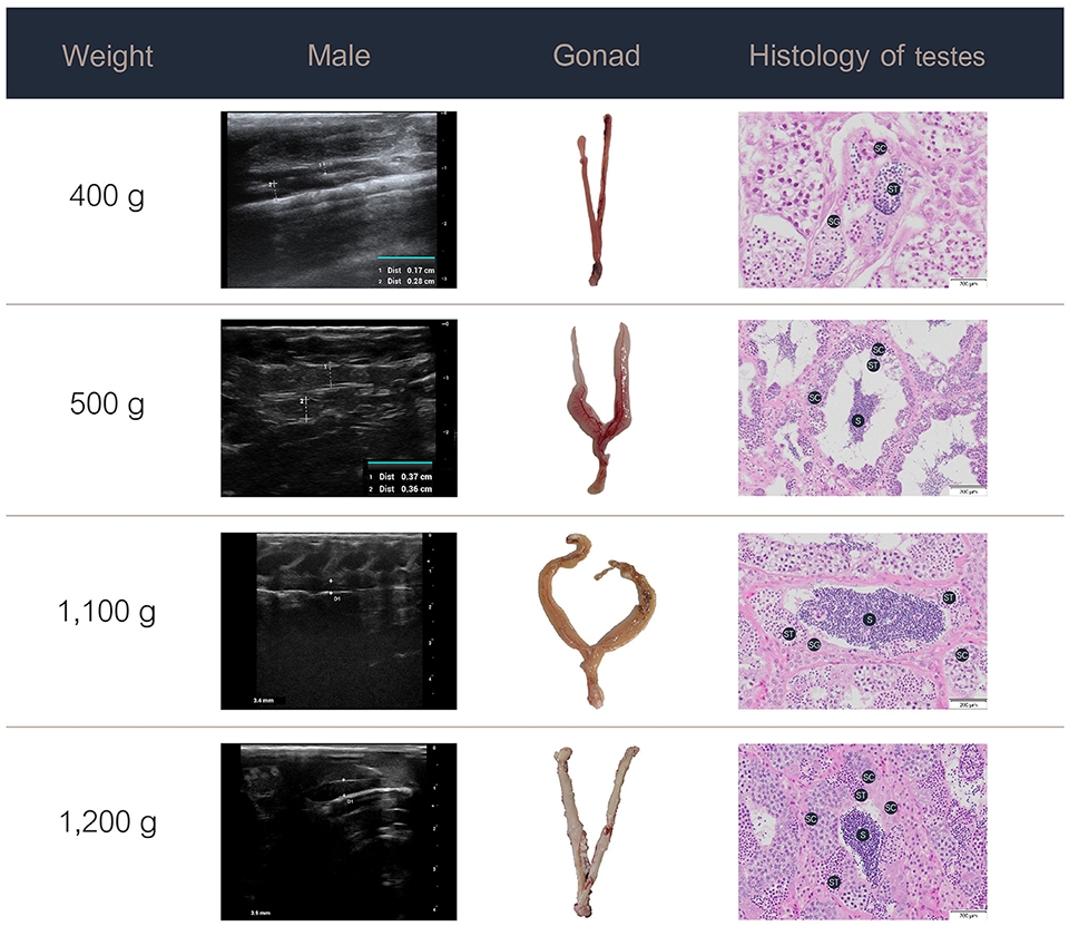
The data on the ultrasound images, gross morphology, and histology of the ovaries in the females and testes in the males at various weight stages are presented in Figs. 3 and 4.
Specifically, the progressive development and maturation of the oocytes correlated with increasing body weight in the female Nile tilapia. At a fish weight of 200 grams, ultrasonographic imaging showed a thin, small ovary measuring approximately 0.35 cm, with histological sections showing oocytes characterized by a small size (50–200 micrometers) and prominent nuclei. For fish weighing 300 grams, the ovary appeared larger (0.84 cm) with more distinct oocyte structures, which included increasing numbers of vitellogenic (process of yolk protein formation in the oocytes during sexual maturation) oocytes (450–750 micrometers) and yolk granules. The female fish weighing 400 grams or more had fully grown ovaries measuring over 1 cm in the longitudinal ultrasound views, which indicated the maturation of ripe-stage oocytes ready for egg release. These mature oocytes were larger (850–1,600 micrometers) than those of the female fish at lower weights and had displaced nuclei and a hydrated yolk.
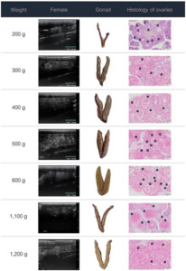
In the male Nile tilapia weighing 400 grams, ultrasound revealed small testes measuring 0.17 cm in the early ripening stage of development. The histological assessment showed spermatogonia, spermatocytes, and limited spermatids in the seminiferous tubules. With increasing body weight of 500 grams and above, the testes measured over 0.4 cm on ultrasound and showed numerous germ cell cysts and wider lumens filled with mature sperm. During the spent stage, the seminiferous tubules contained germ cells and a partially emptied lumen with a reduced number of spermatozoa.
Ultrasonography is a valuable diagnostic tool in veterinary medicine due to its non-invasive technique for assessing reproductive health and sex identification. In this study, we demonstrated the applicability of ultrasonography as a reliable tool for sex identification and reproductive assessment in Nile tilapia. The distinct echogenic patterns observed in the ovaries and testes provided fundamental information to distinguish between the female and male fish, respectively. Ultrasonography presents advantages over traditional techniques, such as manual sex sorting based on external genitalia, which has shown 87 percent accuracy compared to the 95 percent accuracy achieved with ultrasonography and subsequently confirmed by the necropsy results in this study.
In practice in farm operations, the effectiveness of manual sex sorting by examining the external genitalia is highly dependent on the skill and experience of the examiner. Thus, if the examiner lacks sufficient experience, inaccuracies in sex identification can occur, which can complicate broodstock management and fry production. Indeed, combining manual sorting methods with ultrasound could potentially increase the accuracy of sex identification without compromising animal welfare under standard farming conditions. Multiple factors, including fish size, age, maturity stage, and equipment quality, can affect the accuracy of ultrasound interpretations.
The limitations observed in this study, particularly the difficulty in identifying small testes in male Nile tilapia weighing less than 400 grams, highlight the need for improvements in ultrasonographic techniques to enhance accuracy in smaller fish. Adjustments to the frequency and depth settings of the ultrasound machine, as well as improving the sensitivity of the equipment, are crucial. For example, higher frequency sound waves can provide better visualization of internal and reproductive organs in smaller fish. The approach to ultrasound probe positioning is important and varies depending on the anatomy of the fish. Further research should focus on these technological improvements to expand the applicability of ultrasonography across different fish sizes and species.
In terms of applying this technique, future research should focus on using ultrasonographic methods in different ways, for example, to examine the ovary size of fish to determine its correlation with the gonadosomatic index (GSI; the gonad mass as a proportion of the total body mass) and to study ultrasonographic characteristics at different developmental stages. Developing an automated ultrasonography-based system for sex differentiation in fish could facilitate rapid and accurate sex identification, enabling the effective identification of male and female fish. This improvement could benefit commercial breeding operations by improving efficiency, reducing labor costs, and minimizing human error. These approaches have the potential to improve the management of not only Nile tilapia but also other important fish species.
Perspectives
This study demonstrated the applicability of ultrasonography as a non-invasive tool for sex identification and reproductive assessment in Nile tilapia. The echogenic patterns observed in the ultrasound images of the ovaries and testes enabled effective differentiation between the female and male fish, respectively. These findings are consistent with previous research and confirm the application of ultrasound for monitoring gonadal development and reproductive health in tilapia aquaculture.
Importantly, integrating ultrasound technology in fish farming practices has the potential to enhance selective breeding programs, improve production efficiency, and promote sustainable aquaculture. Future research should focus on refining ultrasound techniques and exploring their application in other commercially important fish species to further validate their utility in the industry.
Now that you've reached the end of the article ...
… please consider supporting GSA’s mission to advance responsible seafood practices through education, advocacy and third-party assurances. The Advocate aims to document the evolution of responsible seafood practices and share the expansive knowledge of our vast network of contributors.
By becoming a Global Seafood Alliance member, you’re ensuring that all of the pre-competitive work we do through member benefits, resources and events can continue. Individual membership costs just $50 a year.
Not a GSA member? Join us.
Authors
-

Department of Physiology, Faculty of Veterinary Medicine, Kasetsart University, Bangkok, Thailand
-
Department of Veterinary Microbiology and Immunology, Faculty of Veterinary Medicine, Kasetsart University, Bangkok, Thailand
-
Department of Physiology, Faculty of Veterinary Medicine, Kasetsart University, Bangkok, Thailand
-
Corresponding author
Department of Veterinary Microbiology and Immunology, Faculty of Veterinary Medicine, Kasetsart University, Bangkok, Thailand; and Laboratory of Biotechnology, Chulabhorn Research Institute, Bangkok, Thailand
Related Posts

Intelligence
Bangladesh’s tilapia aquaculture industry shows resilience
Tilapia aquaculture in Bangladesh has developed significantly since 1999, based on the Genetically Improved Farmed Tilapia (GIFT) strain of Nile tilapia (Oreochromis niloticus) introduced from Malaysia and on the significant genetic improvement research work by the Bangladesh Fisheries Research Institute (BFRI).
Health & Welfare
Bioinformatics: The ultimate breeding tool
Bioinformatics, a true hybrid between molecular and computer sciences, changes the rules of both breeding and medicine with a novel approach.
Health & Welfare
‘Super male’ Nile tilapia outperform hormone-reversed fish in Nicaragua
A recent study compared the growth and survival at different salinity levels of “super male” (YY) tilapia with normal sex O. niloticus reversed to 100 percent phenotypic males.
Responsibility
Genetic improvement aids red tilapia growth in Egypt
A new breeding program for genetic improvement of red tilapia was established at the Fish Research Center (FRC), Suez Canal University, in Ismailia, Egypt. It aims to improve the growth rate of the fish and to provide significant benefits to tilapia farmers.