Shrimp necrosis has infectious etiology
Local L. vannamei exhibit unusual cellular immune response
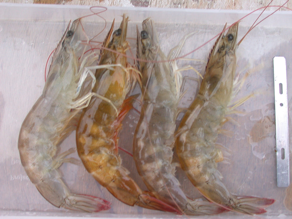
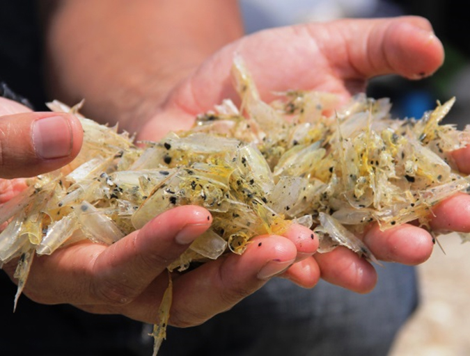
During 2007, Centro Nacional de Acuicultura e Investigaciones Marinas – Escuela Superior Politécnica del Litoral (CENAIM–ESPOL) reported several cases of muscle necrosis in locally farmed Pacific white shrimp (Litopenaeus vannamei). These cases were associated with low mortalities at harvest based on anecdotal evidence from shrimp farmers.
The disease was described as an idiopathic muscle necrosis. It was visually characterized by extensive necrotic areas in striated tail muscle tissues that appeared white and opaque. Furthermore, histological analysis of suspect samples with macroscopic lesions revealed a loss of sarcomeric structure accompanied by coagulative muscle necrosis and hemocytic infiltration.
Muscle necrosis in crustaceans
Histological evidence has suggested two forms of muscle necrosis in crustaceans: one involving extensive hemocytic infiltration and the other a non-inflammatory necrosis in which a weak immune reaction is observed.
Hemocytic infiltration can occur in response to injury caused by pathogens. The authors believe the unusual cellular immune response in local L. vannamei suggested an infectious etiology.
Similar clinical signs have been described for two infectious diseases not locally reported: infectious myonecrosis (IMN) and Penaeid white tail disease (PWTD). IMN affecting cultured L. vannamei in Brazil is caused by a double-stranded RNA virus named infectious myonecrosis virus (IMNV). Meanwhile, PWTD was reported in cultured L. vannamei in Belize. Its etiological agent is Penaeus vannamei nodavirus (PVNV).
CENAIM–ESPOL study
In work by the authors at CENAIM–ESPOL, muscle necrosis was hypothesized to be caused by an infectious agent. To study the nature of the disease, sampling from local shrimp ponds with muscle necrosis cases and challenge tests with cell-free filtrates from diseased shrimp in healthy L. vannamei were performed.
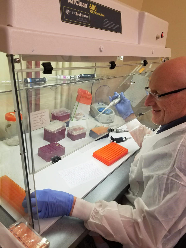
Real-time polymerase chain reaction (RT-PCR) tests for IMNV and PVNV using commercial kits and histological analysis were included for diagnosis. In addition, several samples were sent to the University of Arizona Aquaculture Pathology Laboratory for RT-PCR testing.
The histological exams confirmed most of the challenged shrimp had lesions in skeletal muscle, including multifocal necrosis, fibrocytic inflammation and phagocytosis. Sixty-nine of 99 challenged shrimp showed clinical muscle necrosis.
All challenged shrimp tested by RT-PCR assay were negative for IMNV and PVNV. There were no mortalities throughout the four-week trial. Experimental shrimp in a negative control analyzed by histology showed no signs of muscle necrosis. In addition, there was no evidence of opportunistic bacterial infection in tested tissues from all challenged shrimp.
Perspectives
Interestingly, histological lesions found in the local shrimp were indistinguishable from those reported in L. vannamei with IMN and PWTD. PVNV has not been detected in samples submitted for diagnosis from farms in Central or South America, except from Belize and an adjacent area of Nicaragua.
In the experiments, muscle necrosis could be consistently reproduced through sequential trials, confirming the disease has an infectious etiology. In fact, the results suggested the etiological agent of this disease could be a new infectious agent or a different strain of IMNV.
(Editor’s Note: This article was originally published in the September/October 2012 print edition of the Global Aquaculture Advocate.)
Authors
-

Centro Nacional de Acuicultura e Investigaciones Marinas
Escuela Superior Politécnica del Litoral
Campus Gustavo Galindo Velasco
Km 30.5 Vía Perimetral
P. O. Box 09-01-5863
Guayaquil, Ecuador -
Universidad Estatal Península de Santa Elena
Santa Elena, La Libertad, Ecuador -
Centro Nacional de Acuicultura e Investigaciones Marinas
Escuela Superior Politécnica del Litoral -
Centro Nacional de Acuicultura e Investigaciones Marinas
Escuela Superior Politécnica del Litoral -
Centro Nacional de Acuicultura e Investigaciones Marinas
Escuela Superior Politécnica del Litoral
Related Posts
Health & Welfare
A comprehensive look at the Proficiency Test for farmed shrimp
The University of Arizona Aquaculture Pathology Laboratory has carried out the Proficiency Test (PT) since 2005, with 300-plus diagnostic laboratories participating while improving their capabilities in the diagnosis of several shrimp pathogens.
Responsibility
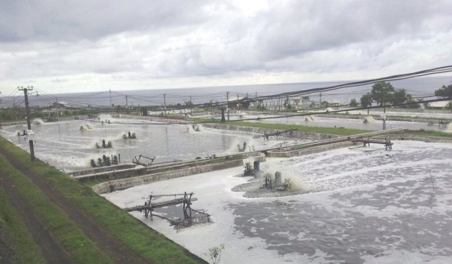
A look at various intensive shrimp farming systems in Asia
The impact of diseases led some Asian shrimp farming countries to develop biofloc and recirculation aquaculture system (RAS) production technologies. Treating incoming water for culture operations and wastewater treatment are biosecurity measures for disease prevention and control.
Responsibility
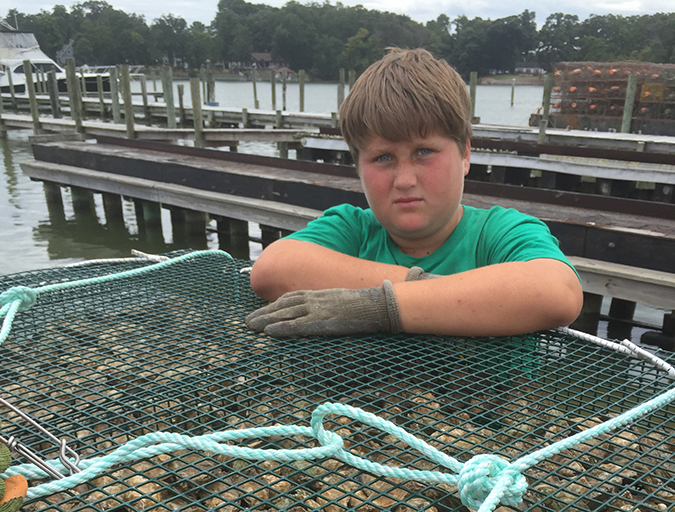
Ailing waterways hail the oyster’s return
The Lower Hudson Estuary and Chesapeake Bay, two waterways once home to thriving oyster beds, would welcome the shellfish’s return. Aquaculture initiatives in both areas aim to reinvigorate the water and the communities they support.
Health & Welfare
A holistic management approach to EMS
Early Mortality Syndrome has devastated farmed shrimp in Asia and Latin America. With better understanding of the pathogen and the development and improvement of novel strategies, shrimp farmers are now able to better manage the disease.