Diseases found in tilapia culture in Latin America
Vibrio isolates have been obtained from intestinal contents of apparently healthy tilapia
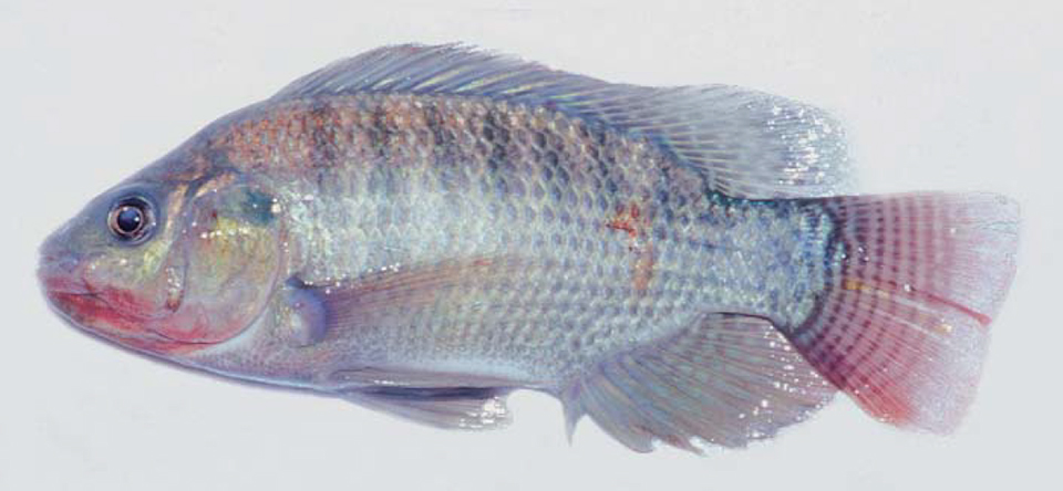
Tilapia tolerate adverse water quality parameters and other stress factors better than many other species used in commercial aquaculture. As a result, tilapia culture has grown worldwide without much implementation of biosecurity measures that have been standardized for less disease-resistant fish like trout and salmon. Currently, several significant diseases affect tilapia. Some are new, and others are old problems that have re-emerged with serious results.
This surge of diseases in tilapias is mainly related to the intensification of culture methods. Every year, tilapia are cultured at higher densities, as well as in recirculating systems, where tilapia grow exceptionally well – but so do their pathogens.
Latin American countries like Costa Rica, Ecuador, and Honduras are the main exporters of fresh tilapia fillets to the United States. The pathogens of tilapia cultured in Latin America fall within three main categories: bacterioses, mycoses, and parasitoses.
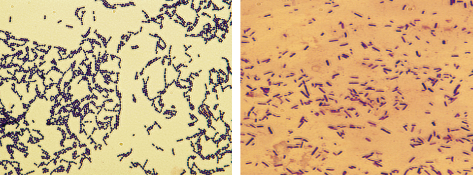
Bacterial diseases
Gram negative bacteria and BHS

![Ad for [Aquademia]](https://www.globalseafood.org/wp-content/uploads/2025/07/aquademia_web2025_1050x125.gif)
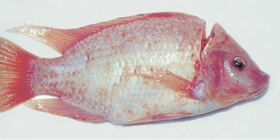
Tilapia affected by gram negative bacteria show signs of darkening, exophthalmia, loss of appetite, and hemorrhagic or ulcerated areas at the bases of the pectoral and ventral fins and in the eye region. Internally, it is common to find pale livers and the presence of hemorrhagic foci. Necrosis of the liver, heart, spleen, and skeletal muscle is often detected, in addition to necrosis of the renal hematopoietic tissue.
Bacterial hemorrhagic septicaemia (BHS) can break out and lead to losses of 5-100 percent in freshwater and brackish waters. Chronic mortalities have been reported in Oreochromis aureus cultured in cages in coastal waters at La Parguera, Puerto Rico, where the salinity was 35 ppt.
In various Latin American countries where tilapia and their hybrids are cultured, bacteria such as Vibrio spp. and Aeromonas hydrophila and other species of motile aeromonads, Edwardsiella tarda, Pseudomonas fluorescens, and other pseudomonads have been isolated from cases of BHS. Isolates of Vibrio spp. have been obtained from the intestinal contents of apparently healthy tilapia in freshwater environments. These fish could represent asymptomatic carriers of the infection. In addition, V. cholerae NON 01 has been isolated from a population of tilapias cultured in the waters of a dam in a Caribbean country. All of these bacteria are normal components of the bacterial flora of tilapia and the aquatic environment in which they are cultured, so they should be considered facultative or opportunistic pathogens that cause disease when the fish are under stress.
Treatment involves administration of antimicrobial substances mixed into feed. It is important to first carry out an antibiotic-sensitivity test with the corresponding bacterial isolate, and then determine the minimum inhibitory concentration (MIC μg per millileter = ppm) to establish the dose rate. These technical procedures should be carried out by qualified personnel, and must take into account the legal norms in force with regard to the use of antimicrobial products in animals destined for human consumption.
Gram positive bacteria
The recent appearance of pathological problems associated with the presence of streptococci in tilapia cultured in several countries in North America, Central America and South America worries producers. One of the streptococci involved – Streptococcus iniae – is currently thought to constitute an emerging public health problem (Weinstein et al., 1997). According to Eldar et al. (1995), S. iniae has a worldwide distribution.
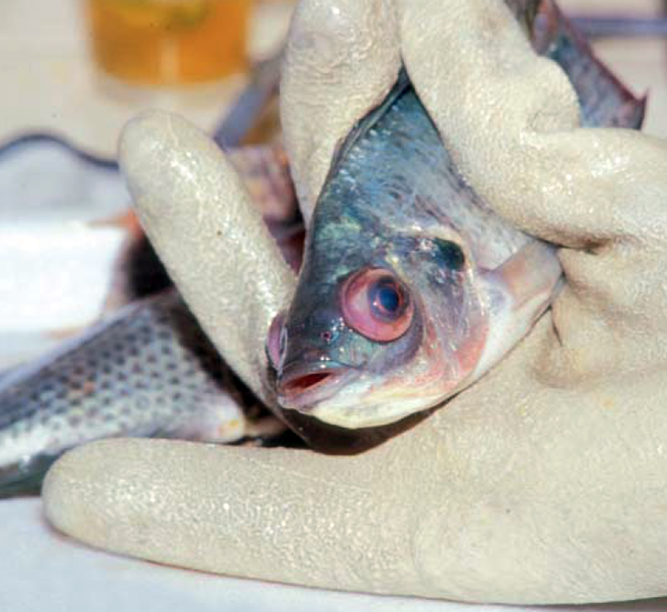
In tilapia, streptococcosis usually produces a chronic disease characterized by the presence of granulomata that affect the spleen, brain, liver, and kidney. The affected tilapia show erratic and disorientated swimming movements as a result of the meningoencephalitis that results. In other cases, the animals show clinical signs similar to those of bacterial hemorrhagic septicaemia.
To date, streptococcosis has been diagnosed in cultured populations of tilapia and their hybrids in two countries of Central America and in two countries of South America.
The prevention of streptococcosis is related to appropriate management and hygiene, including the removal and destruction of affected fish. It is important to precook (pasteurize) any minced fish or fish viscera incorporated into wet feeds for tilapia. The use of antimicrobials to control streptococcosis has been made more difficult by the development of resistant strains. The antimicrobials do not destroy all the bacterial cells stored within the macrophages, which gives rise to the reappearance of the disease once antimicrobial therapy has been discontinued.
Tuberculosis or mycobacteriosis
Very serious problems associated with the presence of mycobacteriosis have been reported in tilapia in some African countries. To date, however, this infection has only been detected in tilapia cultured in two countries of the Central American and region. In one of these instances, Mycobacterium fortuitum was isolated. Mycobacterium chelonae, M. fortuitum and M. marinum are recognized as potential human pathogens and could be transmitted through the inadequate handling of infected tilapia and/or other infected species of fish (Frerichs, 1993).
Diseased fish show small focal granulomata in the liver, spleen, and kidney. The condition could give rise to losses of tilapia fed infected raw fish or viscera of the same. The isolation of mycobacteria is feasible, but very special culture media are required and, in practice, attempts at isolation of the mycobacteria are not always successful.
Columnaris
Columnaris disease is caused in freshwater by the myxobacterium Flexibacter columnaris, which is also known as Chondrococcus columnaris, Cytophaga columnaris and Flavobacterium columnare. Its counterpart in saltwater with salinities higher than 28 ppt is Flexibacter maritimus. These organisms have a wide distribution in tropical, subtropical, and temperate waters with a temperature range of 12.6 to 30 degrees-C. F. columnaris is one of the most commonly encountered bacterial pathogens in tilapia culture, and epizootics normally follow stressful events.
Columnaris is characterized clinically by the presence of greyishwhite, erosive areas or shallow necrotic lesions on the fins, head, or body of fish. The gills are also affected, and usually appear pale and necrotized. Suitable prevention and control measures include maintenance of adequate water quality, population density, and feeding. The use of nifurpirinol and other nitrofuran derivatives has been quite successful in the treatment of columnaris disease.
Studies undertaken in Venezuela have shown promising results regarding the preventive vaccination of “cachamas” (Colossoma macropomum) against columnaris disease in ponds and floating cages (Ravelo, 1994). Therefore, a vaccine may shortly become available to prevent columnaris in cultured tilapia. Experimental vaccines have been developed to prevent infections by Aeromonas hydrophila, Edwardsiella tarda, and streptococcosis in tilapia.
Mycotic diseases
In general terms, dermatomycosis (caused by Saprolegnia spp. and other Phycomycete fungi) is considered a secondary infection related to inadequate hygiene or farm management. Following capture and transfer of tilapia from one pond to another, up to 50 percent of the fish can be affected by dermatomycosis.
The disease manifests itself by the presence of lesions on the fins, mouth, and skin. These lesions are covered by a white or greyish-white cottony mass, the fungal mycelium. The infection is also extremely common in dead eggs, from where it can spread to live eggs and cause their death through asphyxia. Dermatomycosis is frequently associated with a simultaneous bacterial infection.
Principal parasitic diseases
In Latin America, many parasites are commonly found in tilapia in both natural environments and under culture conditions. Some of these parasites (e.g., some of the protozoans and monogeneans) are of particular significance in the process of sex reversal, where their presence can result in heavy mortalities or, at the very least, weakened fish.
Ciliates
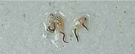
Ichthyophthirius multifiliis, commonly known as “white spot” or “itch,” is a relatively large parasite capable of causing serious losses in tilapia cultured in freshwater. It produces lesions on the skin, gills, pharynx, and nares. The entire surface of the fish – particularly in the case of larvae – is affected within 24 hours of initial infection. The ideal temperature for itch propagation is around 24 degrees-C. It is interesting to mention that Sin et al. (1994) and Subasinghe and Sommerville (1989) have demonstrated that female Oreochromis aureus and O. mossambicus vaccinated with live teronts of I. multifiliis transmitted a passive immunity against ichthyophthiriasis to their larvae.
Chilodonella sp. has been observed as a parasite of the skin and gills of cultured tilapia, especially in the case of larvae undergoing sex reversal. It can be of considerable importance with stressed fish.
The trichodinids, which include the genera Trichodina, Trichodinella, and Tripartiella, are peritrichous ciliates normally found on the gills and skin. They are an almost natural but troublesome companion during the sex-reversal process. Their presence is indicative of high population densities and poor water quality. Trichodina spp. is a very special problem in mouth-rearing tilapias, as it invades the mouth and transmits the infection to the larvae.
Flagellates
Ichthyobodo necator, or Costia necatrix, is a parasite with great pathological significance in tilapia larvae, particularly under high fish densities and for stressed fish. I. necator generally occurs on the gills, and its detection is quite often difficult because of its small size. Affected fish show signs of general weakness, with their fins held close to the body. There is opacity of the skin, and the gills are hyperplasic with abundant mucus production.
Amyloodinium ocellatum is a saltwater parasite. In smaller fish, the infection seems to be limited to the skin, whereas in larger fish it becomes established on the gill filaments and in the integument of the bucco-pharyngeal region. Its control involves baths of copper sulphate pentahydrate or changes in water salinity.
Piscinoodinium pillulare is a parasite usually found on the skin and gill filaments, but it is capable of penetrating beneath the skin epithelium and establishing itself in the subcutaneous tissue. It is an important emerging pathological problem in tilapia culture operations.
Treatment for most of these protozoan infections typically involves formalin baths.
Monogeneans
The monogeneans are parasites that have an exfoliating action on the skin and gills of fish. They feed on the mucus and epithelium of the body surface, producing external lesions that erode and expose the dermis to infections by viruses, bacteria, and fungi. As in the case of trichodinids, monogeneans are considered “almost natural companions,” but they are extremely aggressive organisms.
Monogeneans are often present in the sex-reversal process, where their infestation is favored by their direct life cycle in high fish densities. They can be controlled by baths in formalin, potassium permanganate, hydrogen peroxide, or organo-phosphorus compounds, or by salinity changes repeated as often as necessary. These parasites (e.g., Gyrodactylus spp. and Cichlidogyrus spp.) can cause heavy mortalities in tilapia within a short period of time.
Neobenedenia melleni is of particular importance in tilapia culture, as it has eliminated entire populations of tilapia cultured in brackish and coastal marine waters in the Bahamas, Jamaica, Puerto Rico, and other areas of the Caribbean (Bunkley-Williams & Williams, 1995). It must be emphasized that tilapia, as exotic species in the region, do not have resistance to this monogenean, so they tend to die very rapidly once the infection has become established.
Information currently available suggests that Neobenedenia melleni can be controlled with water baths (18 ppt for seven consecutive days, to be repeated after three months) or baths using formalin, potassium permanganate, or organo-phosphorus compounds. These measures are extremely costly and difficult to apply in commercial tilapia culture operations. When tilapia are cultured in floating cages, infections can be avoided by moving the cages to deeper water areas.
Digeneans
Metacercariae of the diplostomatid Diplostomum compactum produce a condition known as “eye fluke,” “cataract,” or “parasitic blindness” in tilapia, as well as native species of cichlids in several countries of Central and South America. Under high infection levels, the unpleasant appearance of affected fish makes consumers reject the product.
Crustaceans
In Peru, a serious epizootic caused by the copepod Caligus sp. has been reported in tilapia hybrids cultured in brackish water (Burmeister, 2000: personal communication). Affected fish showed signs of erosion and hemorrhages of the skin surface. The infection was controlled using freshwater baths.
Other pathological problems
Recent research in Venezuela (Conroy, 2000) has shown that aflatoxicosis is potentially a very significant problem in commercial tilapiaculture operations. The condition is due to the utilization of pelleted feeds contaminated with aflatoxin-producing molds.
Affected fish show patent growth variations, together with various types of macroscopic and microscopic changes associated with the presence of aflatoxins. This is a problem that must be immediately addressed both by feed producers and tilapia farmers to guarantee the aflatoxin-free status of the final product offered to consumer markets.
Note: Cited references are available from the author.
(Editor’s Note: This article was originally published in the December 2001 print edition of the Global Aquaculture Advocate.)
Now that you've reached the end of the article ...
… please consider supporting GSA’s mission to advance responsible seafood practices through education, advocacy and third-party assurances. The Advocate aims to document the evolution of responsible seafood practices and share the expansive knowledge of our vast network of contributors.
By becoming a Global Seafood Alliance member, you’re ensuring that all of the pre-competitive work we do through member benefits, resources and events can continue. Individual membership costs just $50 a year.
Not a GSA member? Join us.
Author
-

Pharma-Fish S.R.L.
Apartado de Correo No. 406
Maracay 2101-A
Estado Aragua, Venezuela
Related Posts
Health & Welfare
Streptococcosis in tilapia
Tilapia infected with streptococcosis pathogens exhibit similar clinical signs, including lethargy, abnormal swimming and lack of appetite.
Health & Welfare
Health management of barramundi
By combining husbandry practices aimed at stress reduction with better disease management higher barramundi survival rates will be the likely result.
Health & Welfare
Parasite treatment reduces F. columnare infection in tilapia
The authors conducted a study to evaluate whether treatment of Trichodina-parasitized tilapia with formalin would improve fish survival and reduce F. columnare infection. Tilapia not treated with formalin showed significantly higher mortality than treated fish.
Health & Welfare
Developing live bacterial vaccines by selecting resistance to antibacterials
As wide use of antibiotics has led to antibiotic resistance in fish pathogens, vaccines present an alternative control method to prevent bacterial diseases.