A study of Zoea-2 Syndrome in hatcheries in India, part 1
Overview and methodology for examination of Pacific white shrimp mortalities
Shrimp aquaculture in India is a highly dynamic and fast-growing activity dominated by the exotic Pacific white shrimp (Penaeus vannamei), introduced in 2009. The success of the commercial shrimp industry depends mainly on the availability of healthy and quality seedstock, and 276 shrimp hatcheries are involved in the production of specific pathogen free (SPF) P. vannamei seedstock and serving the shrimp aquaculture sector.
Because of the constant demand of the growing aquaculture sector, intensive shrimp farming has improved substantially during the decade. This increasing trend of intensification and commercialization has exacerbated disease epizootics.
Diseases are significant challenges for shrimp larval breeding systems, particularly bacterial diseases such as luminescent bacterial diseases that cause severe economic consequences for hatchery operations. Large-scale losses of eggs and larvae have been reported due to larval mycosis caused by Legenidium spp. and Sirolpidium spp. Larval fouling is caused by protozoans such as Zoothamnium and Vorticella, in addition to other diseases of viral and bacterial origin.
However, since introducing Vannamei, Indian shrimp hatcheries have experienced larval mortality in the zoea-2 stage, with molt deterioration and resulting in heavy mortality. In the case of P. vannamei shrimp larvae in Ecuador, Mexico and the United States, similar larval losses were recorded with the “zoea-2 syndrome” in 1993.
The occurrence of mortalities in zoeal stages of P. vannamei frequently reported in Indian shrimp hatcheries provided us with the stimulus to investigate the problem holistically, considering the possible participation of biotic and abiotic factors. This article is adapted from Aqua Cultura (Ecuador), #119, September 2017. We acknowledge Prof. Pushpa Viswanathan of the Electronic Microscopy Transmission Department of the Chennai Cancer Institute for his kind help and support in TEM studies, and also thank the shrimp hatcheries in Tamil Nadu and Andhra Pradesh for contributing samples and information for this study; and acknowledge the International Committee of Animal Registries (ICAR) of India for financial support to carry out this work.
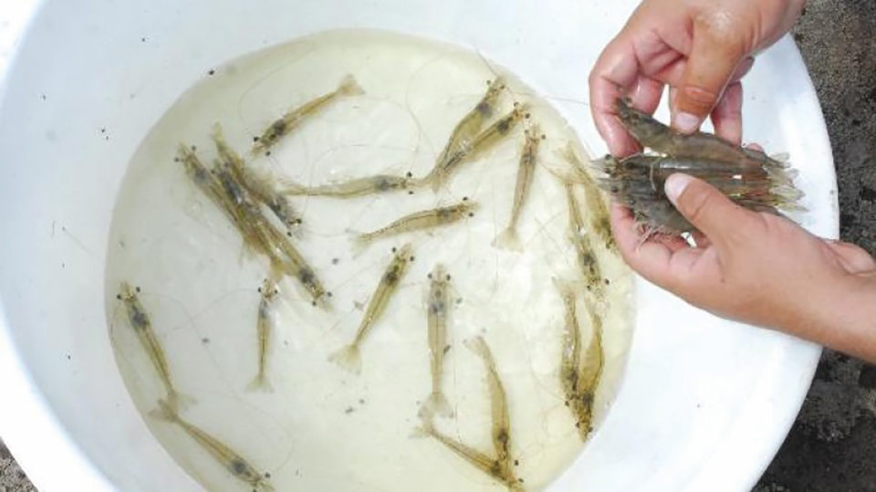
We investigated mortalities of Pacific white shrimp (Penaeus vannamei), during the zoea stages in 15 shrimp hatcheries located on the east coast of India. This disease, commonly known as the zoea-2 syndrome, is characterized by a reduction in the feeding rate of late zoea-1 and early zoea-2 larvae, due to the alteration in their metamorphosis followed by high mortalities. Microscopic studies revealed systemic abnormalities in affected larvae as well as pathological manifestations in the hepatopancreas and intestine. Microbiological detection revealed the predominance of Vibrio alginolyticus in most hatcheries (nine) affected by zoea-2 syndrome.
Histological examination in the hepatopancreas and intestine revealed vacuolization, in addition to detachment of epithelial cells and disintegration of the peritrophic membrane of the intestinal epithelium. The OIE (World Organisation for Animal Health) polymerase chain reaction (PCR) protocols confirmed that the OIE-listed shrimp viral pathogens were absent in the affected larvae. Ultrastructural observation of the pathological manifestation in the hepatopancreas and intestines could not reveal the presence of pathogens. Data on water quality parameters were in the normal range and did not appear to influence the outbreak of zoea-2 syndrome in the nursery sites.
The feeding schemes (with microalgae Skeletonema, Chaetoceros, Thalassia) were uniform throughout the larval cycles, and it was also found that they were not associated with the zoea-2 syndrome. Pathological manifestations of the hepatopancreas and intestine further indicated a deterioration of the digestion and absorption capacity, which resulted in delayed molting and the subsequent death of larvae in a gradual and progressive manner, with a cumulative mortality of 30 to 100 percent in the zoea-2 stage. It was also determined that the continuous storage of nauplii for three to four days within the same larval incubation unit exacerbated the incidence in the nine affected nurseries (OR-48, IC 2.5-932.9). The etiology (cause of disease) of the zoea syndrome may not be due to known infectious agents, an additional result from the interpretation of our studies.
Methodology: Sampling and microbiological exam
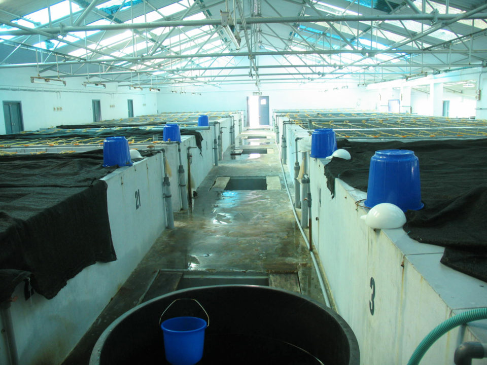
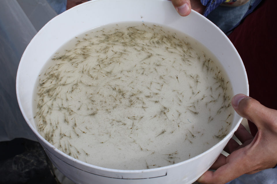
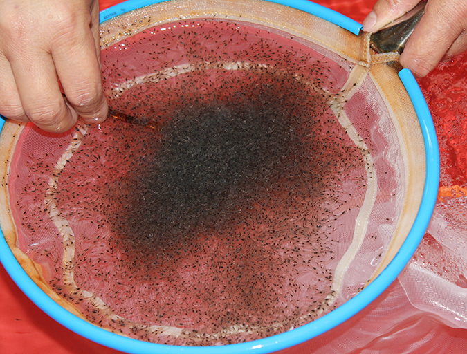
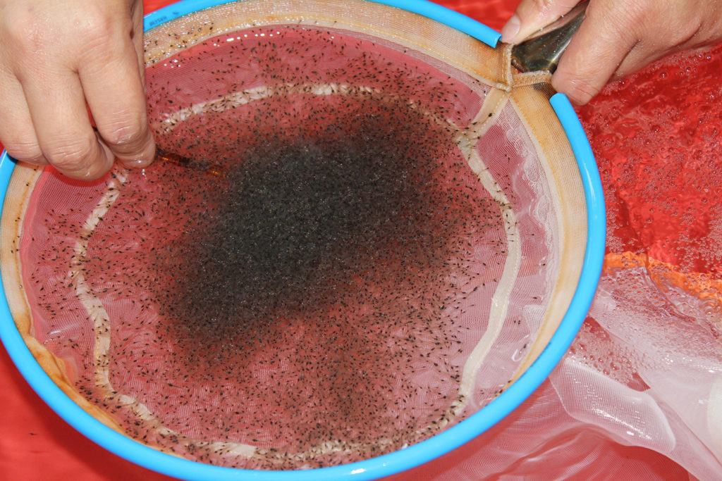
The study involved 15 commercial shrimp hatcheries along the eastern coast of India, including six from Tamil Nadu (Kancheepuram and Vilupuram districts) and nine from Andhra Pradesh (Nellore, Prakasham and East Godavari districts). Larval samples were collected under live conditions from larval cycles affected by zoea syndrome. Live zoeas were observed under optical microscopy and samples of larvae conserved in Davidson AFA fixative for histology. Zoeal samples were fixed in 2.5 percent glutaraldehyde and 0.1 M sodium cacodylate buffer for electron microscopic observations. The larvae were also conserved in 90 percent ethanol and RNAlater (Ambion) for the detection of pathogens by PCR analysis.
During the microbiological examination, samples of 0.5-gram larvae were collected and placed in sterile phosphate buffered saline (PBS) solution, homogenized and inoculated on thiosulfate bile salt sucrose (TCBS) agar and Zobell marine agar (ZMA). The total plate count (TPC) was performed by means of 10-fold serial plate dilution plates in duplicate on ZMA. Plates were incubated at 30 ± 1 degrees-C and observed after 24 hours. Based on phenotypic characteristics, pure cultures of the dominant heterotrophic bacterial flora were identified.
DNA extraction, RNA extraction and cDNA synthesis
Genomic DNA was extracted from larval samples, as described in the experiment by Rajendran et al. (2016). Larval samples were homogenized and digested for 10 minutes at 95 degrees-C in 500 μl of lysis buffer (50 mM Tris, 1 mM methylenediaminetetraacetic acid (EDTA), 500 mM NaCl 1 percent SDS) and 0.1 mg Proteinase K. The mixture was centrifuged at 12,000 rpm for 10 minutes at 4 degrees-C. After centrifugation, the supernatant was carefully collected, and two volumes of ethanol were added and maintained at minu-20 degrees-C for one hour. The mixture was then centrifuged at 12,000 rpm for 10 minutes at 4 degrees-C. The DNA sediment was washed with 70 percent cold ethanol, air dried, re-suspended in nuclease-free water and stored at minus-20 degrees-C.
RNA was extracted from larval samples using TRIzol ™ Reagent following the manufacturer’s protocol. The amount and quality of the extracted RNA were evaluated using a nano spectrophotometer and stored at minus-80 degrees-C. Reverse transcription was carried out using the iScript cDNA synthesis kit in 10 μl reactions, according to the manufacturer’s instructions and the cDNA was stored at minus-20 degrees-C until further use.
Detection of viral pathogens
Nucleic acids were used for the detection of viral pathogens, extracted from samples of larvae by PCR. For the detection of White Spot Syndrome Virus (WSSV), we used a nested PCR protocol.
Other DNA and RNA viruses – namely, hypodermic and hematopoietic infectious necrosis (IHHNV), baculovirus monodon (MBV), hepatopancreatic virus (HPV), yellowhead virus (YHV), Taura syndrome virus (TSV) and Infectious myonecrosis (IMNV) – were detected by the PCR tests recommended by the OIE. Covert mortality syndrome virus (CMNV) was tested by a nested RT PCR protocol, described by Zhang et al. (2014). PCR was carried out in a thermocycler. An aliquot of the amplified PCR product was resolved on 2.0 percent agarose-Tris-acetate-EDTA (TAE) gels stained with 0.5 μg/mL of ethidium bromide, and the amplified DNA along with a marker of 100 bp DNA was visualized under UV illumination using a gel documentation system (Bio-Rad Laboratories, USA).
Light microscopy exam
The larval samples collected under live conditions of normal and affected larval cycles were observed under light microscopy. For histology, the zoeal samples were fixed in the Davidson AFA fixative for 48 hours and processed using routine histological techniques. The zoeal samples were dehydrated through graded alcohols (70, 90 and 100 percent, respectively) each for 60 minutes. After dehydration, the tissues were cleared twice with xylene for 60 minutes, further infiltrated with paraffin wax for 2 hours and then blocks were prepared using Leica EG 1160 tissue. Tissue sections 4-5 μm thick were obtained on clean microscopic slides of paraffin blocks using a Leica RM 2145 microtome, and the sections subsequently treated by hematoxylin and eosin staining using standard procedure. The stained tissue sections were mounted on DPX and observed under a microscope.
Electron transmission microscope
The larval samples were fixed in 2.5 percent glutaraldehyde in 0.1 M sodium cacodylate buffer (pH 7.3) for 8 hours at 8 degrees-C, fixed in 0.1 percent osmium tetroxide prepared, using the same buffer at 8 degrees-C for 2 hours and processed according to the protocols of Naveenkumar et al. (2013). Sections were examined using a JEM 1400 Transmission Electron Microscope at an accelerated 80 kV voltage and photomicrographs were taken at the Cancer Research Institute (WIA) Adyar, Chennai.
Statistical and epidemiological analyses
The odds coefficient, which measures the strength of the association between disease and exposure to a risk factor, was estimated using the Epi Info ™ 7.1.2 single table analysis at a 95 percent confidence level, by approximation of the Taylor series, and two p-values using the Fisher exact test.
Authors
-

ICAR-Central Institute of Brackishwater Aquaculture
#75 Santhome High Road, Raja Annamalai Puram
Chennai 600028, India -
ICAR-Central Institute of Brackishwater Aquaculture
#75 Santhome High Road, Raja Annamalai Puram
Chennai 600028, India -
ICAR-Central Institute of Brackishwater Aquaculture
#75 Santhome High Road, Raja Annamalai Puram
Chennai 600028, India -
ICAR-Central Institute of Brackishwater Aquaculture
#75 Santhome High Road, Raja Annamalai Puram
Chennai 600028, India
-
ICAR-Central Institute of Brackishwater Aquaculture
#75 Santhome High Road, Raja Annamalai Puram
Chennai 600028, India
Related Posts
Health & Welfare
Acclimating shrimp postlarvae before pond stocking
Shrimp postlarvae acclimation before stocking into the various growout systems (ponds, raceways, tanks) is a critical – and often overlooked, sometimes taken for granted – step in the shrimp culture process. Various water quality parameters should be changed slowly so that the young shrimp have the time to gradually adapt to the new conditions.

Health & Welfare
Born in Hawaii, SPF broodstock shrimp industry faces globalization
The next step for shrimp breeding will be developing animals that aren’t just disease-free, but increasingly resistant to multiple pathogens. The industry is globalizing, with suppliers setting up shop overseas. But its birthplace will always be Hawaii.

Aquafeeds
Biofloc consumption by Pacific white shrimp postlarvae
The stable isotopes technique with δ13C and δ15N can be used to determine the relevance of different food sources to shrimp feeding during the pre-nursery phase of Litopenaeus vannamei culture. During this trial, different types of commercial feed, microalgae, Artemia sp. nauplii and bioflocs were used as food sources.
Health & Welfare
How good are your shrimp postlarvae?
Stocking the best quality shrimp postlarvae, healthy and free of pathogens, is a critical management step with significant effects on the production and profitability of a shrimp farm.