Lipid oxidation results from heme catalysis
Rancid flavor and changes in color, texture and nutritional value are consequences
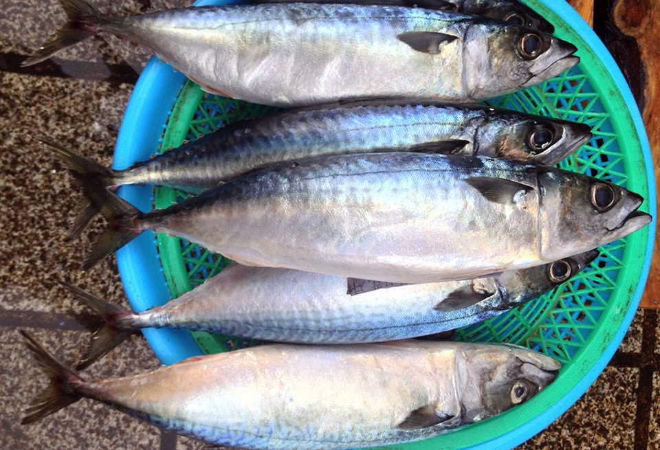
For many years, the roles of dietary eicosapentaeonic acid and docosahexaenoic acid have been related to improved cardiovascular health and brain development in humans. The high degree of these unsaturated fatty acids in fish makes them a good source of the nutrients, but the close proximity of the fatty acids to strong pro-oxidative systems predisposes them to oxidation, which converts them into compounds that negatively affect the quality attributes of the fish.
Rancid flavor is a well-known consequence of lipid oxidation, but changes in color, texture and nutritional value can also develop. The extent of undesirable changes in fish lipids is due to the types of compounds participating in the oxidation process.
Pro-oxidants
Most well known among the pro-oxidants in fish are the transition metals, such as iron and copper, which impact several steps of the oxidation chain. These metals are most active in their reduced states.
A large part of the iron in fish muscle is bound in hemoproteins such as myoglobin, hemoglobin and cytochromes. The physiological functions of myoglobin and hemoglobin are to carry and distribute oxygen to the different tissues. Hemoglobin is the main pigment in red blood cells, and myoglobin is the main pigment in muscle cells. Both myoglobin and hemoglobin have catalytic properties due to their ability to break down hydroperoxides and their suggested activation by hydrogen peroxide into the highly reactive porphyrin cationic radical.
About two-thirds of body iron is found in hemoglobin, and smaller amounts are in myoglobin. A very small amount comprises components in various iron-containing enzymes and in the transport protein transferrin. The remainder is present in the intracellular storage proteins ferritin and hemosiderine. A small pool of non-protein non-heme iron provides “free” iron at micromolar concentrations in tissues.
Metals and heme compounds
Both non-heme iron and hemoproteins can function as pro-oxidants when in contact with pure lipids. However, the situation with muscle is much more complex. Inorganic iron is a strong catalyst in mackerel meat lipid oxidation, whereas heme iron is the major catalyst of lipid oxidation in mullet flesh.
The oxidative rancidity in cooked meats has been attributed to both heme and non-heme iron. It has been further reported that the increased rate of lipid oxidation in cooked meats is due to the release of non-heme iron during cooking. Heme pigments may be more active lipid oxidation catalysts with iron in the ferric state, whereas non-heme iron appears to be more active in the ferrous state. Research has shown that in raw meat and model emulsions, ferric hematin pigments are powerful catalysts of lipid oxidation, whereas in heated meats, the system is more complex, and inorganic iron may play a more important role.
Heme proteins have been considered catalyzers of the propagation step and not true initiators of lipid peroxidation. It has been reported that active species formed by the interaction of hydrogen peroxide with metmyoglobin or methemoglobin, the respective oxidized analogues of myoglobin and hemoglobin, could be described as true initiators of lipid peroxidation. Microsomal oxidase systems coupled with ferric or ferrous iron may also be initiators of lipid oxidation.
The loss of redness in meat is an indicator that lipid oxidation processes mediated by hemoglobin are progressing. Just after death, hemoglobin in muscle tissue is primarily in a reduced state in which, for example, the mixture of oxyhemoglobin and deoxyhemoglobin possesses a red color.
With increased postmortem aging, this reduced hemoglobin auto-oxidizes to methemoglobin, a brown pigment. Methemoglobin is considered more pro-oxidative than reduced hemoglobin due to its less tightly bound heme group and reactivity with hydrogen peroxide and lipid peroxides to form hypervalent hemoglobin catalysts.
Pro-oxidative properties
Numerous studies have shown that certain hemoglobins promote lipoid oxidation more effectively than hemoglobins from other species. For example, herring and mackerel hemoglobins oxidized washed cod lipids more effectively than trout hemoglobins did. Oxygen affinity appeared to play a role, in that those hemoglobins with elevated deoxyhemoglobin contents at pH 6.3 (for example, trout hemoglobin) promoted lipid oxidation most effectively at pH 6.3.
Hemoglobins from pollock, mackerel, menhaden and flounder were found to be equally pro-oxidative at pH 6.0, while they differed significantly in their ability to oxidize cod membrane lipids at pH 7.2. The higher activities at pH 6.0 could be explained by higher and more rapid formation of deoxy- and methemoglobin.
Autoxidation in cold-adapted fish was found to be 10-fold faster than in warmwater fish at all temperatures. The rate of autoxidation was lower in monomeric hemoglobins from, for example, hagfish and lamprey than in tetrameric hemoglobins from carp and tuna.
Blood and lipid oxidation
There is a wide variation in the amounts of hemoglobin extracted from the muscle tissue of bled and unbled fish (Table 1). Myoglobin content was minimal when compared to the hemoglobin content in mackerel light muscle and trout whole muscle. Hemoglobin made up to 65 and 56 percent, respectively, of the total heme protein by weight in dark muscle from unbled and bled mackerel.
Flick, Hemoglobin and myoglobin concentrations, Table 1
| Sample | Hemoglobin (µmol/kg/ tissue) | Myoglobin (µmol/kg/ tissue) | Myoblobin In Total Heme Protein (%) |
|---|---|---|---|
| Trout whole muscle, unbled | 11.10 ± 4.95 | N.D. | N.D. |
| Trout whole muscle, bled | 7.39 ± 2.93a | N.D. | N.D. |
| Mackerel light muscle, unbled | 6.07 ± 1.02 | N.D. | N.D. |
| Mackerel light muscle, bled | 3.40 ± 0.48b | N.D. | N.D. |
| Mackerel dark muscle, unbled | 158.80 ± 21.00 | 342.00 | 35 |
| Mackerel dark muscle, bled | 121.80 ± 17.00 | 382.80 | 44 |
Bleeding significantly reduced rancidity in minced trout whole muscle, minced mackerel light muscle and intact mackerel dark muscle, but not minced dark mackerel stored at 2 degrees C. It was suggested that blood-mediated lipid oxidation in fish muscle depends on various factors that include hemoglobin concentration and type, plasma volume and erythrocyte integrity.
Lipid oxidation in slices of bled and unbled Asian seabass during 15 days of ice storage showed that bled samples had lower peroxide values and thiobarbituric acid-reactive substances throughout the storage period (P < 0.05). Bleeding effectively lowered the total heme and non-heme iron contents of the samples.
The release of non-heme iron was pronounced in the unbled samples during storage. The level of heptanal, the major volatile compound detected in the unbled samples, was four-fold higher than in bled counterparts. The contents of aldehydic compounds, including hexanal, octanal, nonanal and nonenal, were also higher in the former samples.
(Editor’s Note: This article was originally published in the January/February 2014 print edition of the Global Aquaculture Advocate.)
Authors
-

University Distinguished Professor
Food Science and
Technology Department
Center for Applied Health Sciences
Duck Pond Drive
Virginia Tech (0418)
Blacksburg, Virginia 24061 USA -

Assistant Professor
Food Science and
Technology Department
Center for Applied Health Sciences
Duck Pond Drive
Virginia Tech (0418)
Blacksburg, Virginia 24061 USA
Related Posts

Intelligence
Off the Knife with Ned Bell, Vancouver Aquarium
We delve deeper into the foodservice industry’s engagement with sustainable seafood and aquaculture by talking to Ned Bell, the Vancouver Aquarium’s new executive chef. He continues to call on his Canadian culinary comrades to learn more about seafood, then help educate consumers.
Aquafeeds
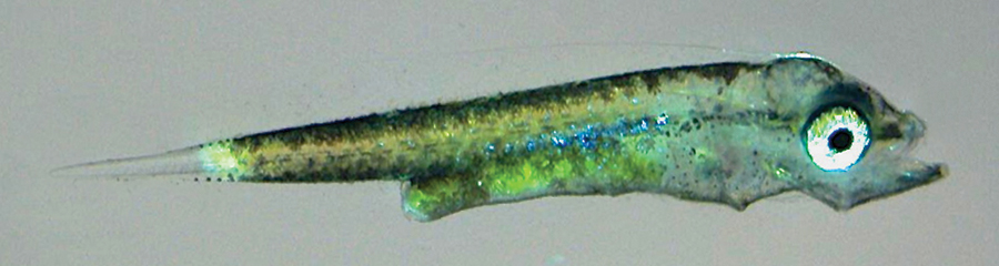
Liposomes open new doors in larval fish nutrition
Liposomes, synthetic microparticles originally developed for the pharmaceutical industry, can be produced for ingestion by rotifers and artemia. A study found that yellowtail larvae fed rotifers enhanced with liposomes containing taurine had improved growth.
Aquafeeds
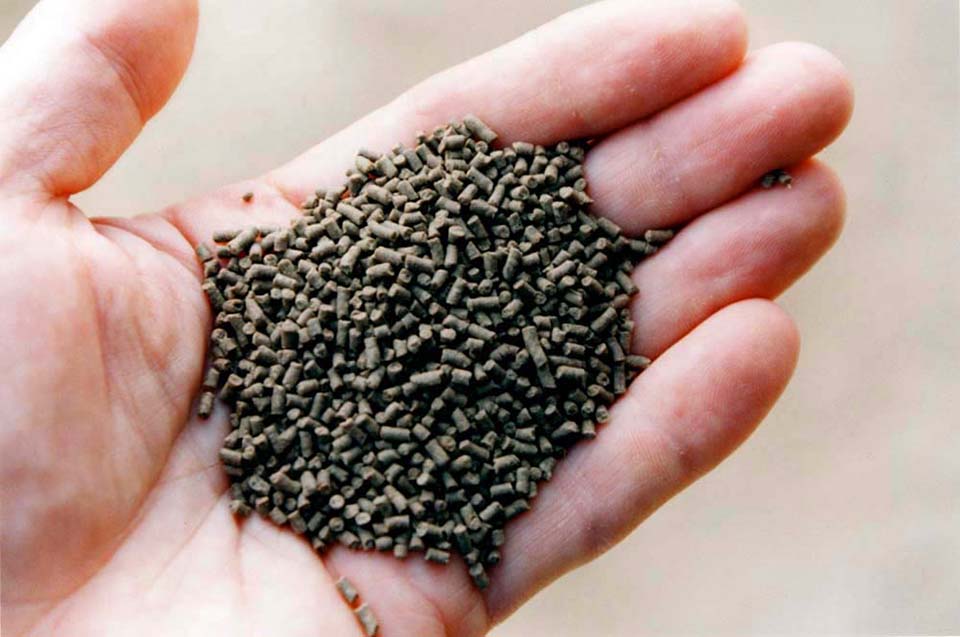
Hidden value of feed: Water quality
The intrinsic values of better feeds directly affect shrimp growth and performance in a positive way, but because better feeds produce less waste materials in culture systems, they also result in improved water quality that indirectly improves shrimp performance.
Aquafeeds
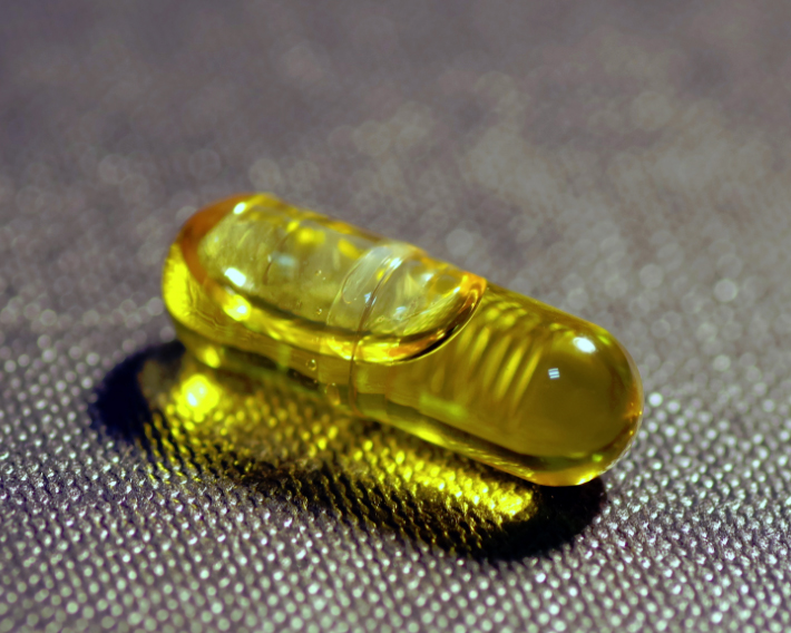
Considerations for alternative ingredients in aquafeeds
A key to expanding aquaculture is finding alternative sources of proteins and oils. Supplementing or replacing fish oil in aquaculture feeds with alternative lipid sources – oils seeds, microalgae, insects and others – appears possible as long as essential fatty acid (EFA) requirements are satisfied.