Early Mortality Syndrome affects shrimp in Asia
Both black tiger and Pacific white shrimp affected by disease
A new disease appeared in shrimp farms located in southern China and Hainan Island in 2010. By early 2011, “Early Mortality Syndrome” (EMS) was also detected in Vietnam and Malaysia.
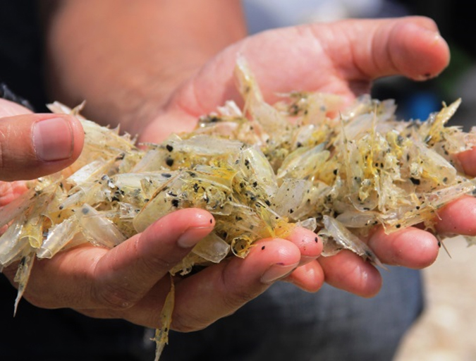
The disease appears within 20 to 30 days of stocking ponds with postlarvae. Both black tiger shrimp (Penaeus monodon) and Pacific white shrimp (Litopenaeus vannamei) are affected by the disease.
Mortalities can approach 100 percent in severely affected ponds, where diseased shrimp become lethargic and anorexic. Upon simple dissection, the hepatopancreas organs of the shrimp may appear atrophied and whitish with black streaks. Other signs include a soft, generally darker shell and mottling of the carapace.
Pathology
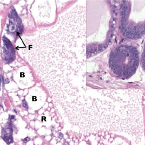
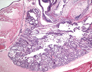
Both P. monodon and L. vannamei with EMS present the same pathology. Samples of shrimp preserved for histology have shown the effects of EMS appear to be limited to the hepatopancreas (H.P.). Progressive dysfunction of the H.P. results from lesions that reflect degeneration and dysfunction of the tubule epithelial cells that progress from proximal to distal.
The first changes observed in the hepatopancreases of affected shrimp is a marked reduction of fat storage cell vesicles and loss of oil/fat droplets, as well as a decrease in the activity of secretory cells. As the disease progresses, fat, basophilic and secretory cells degenerate and begin to round up, detach from the H.P. tubule basement membrane and slough into the H.P. tubule lumen.
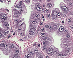
Concomitant with the degeneration of these cells in the more proximal regions of the H.P. tubules, the number of mitotically active E cells declines, and the marked inflammatory response is dominated by hemocyte infiltration and encapsulation of the affected H.P. tubules.
As the tubule epithelial cells degenerate, their nuclei become variably hypertrophic, and the nuclei of most nuclei become enlarged. In the terminal stages of the disease, a severe secondary infection likely caused by opportunistic Vibrio bacteria occurs in the sloughed masses of epithelial cells in the H.P. tubule lumens. Affected shrimp die from H.P. dysfunction and the terminal vibrio infection.
Etiology
This degenerative pathology of the hepatopancreas is highly suggestive of a toxic etiology. Similar lesions have been reported in the H.P.s of shrimp exposed to aflatoxin B1 and the mitosis inhibitor benomyl, which supports this theory.
Studies to determine the etiology of EMS run at the University of Arizona Aquaculture Pathology Laboratory have not been successful. The laboratory has tested commercial feeds collected at shrimp farms with EMS, and frozen samples of shrimp with EMS from affected farms were used in infectivity studies. A crustacide commonly used in the region to kill vectors of white spot syndrome prior to stocking has also been tested.
To date, the University of Arizona lab has not experimentally induced lesions of the hepatopancreas consistent with those observed in shrimp with EMS.
(Editor’s Note: This article was originally published in the January/February 2012 print edition of the Global Aquaculture Advocate.)
Authors
-

OIE Reference Laboratory for Shrimp Diseases
Department of Veterinary Science and Microbiology
University of Arizona
Tucson, Arizona 85721 USA -

Department of Veterinary Science and Microbiology
University of Arizona -
Department of Veterinary Science and Microbiology
University of Arizona -
Department of Veterinary Science and Microbiology
University of Arizona -
Department of Veterinary Science and Microbiology
University of Arizona
Related Posts
Health & Welfare
A holistic management approach to EMS
Early Mortality Syndrome has devastated farmed shrimp in Asia and Latin America. With better understanding of the pathogen and the development and improvement of novel strategies, shrimp farmers are now able to better manage the disease.
Responsibility
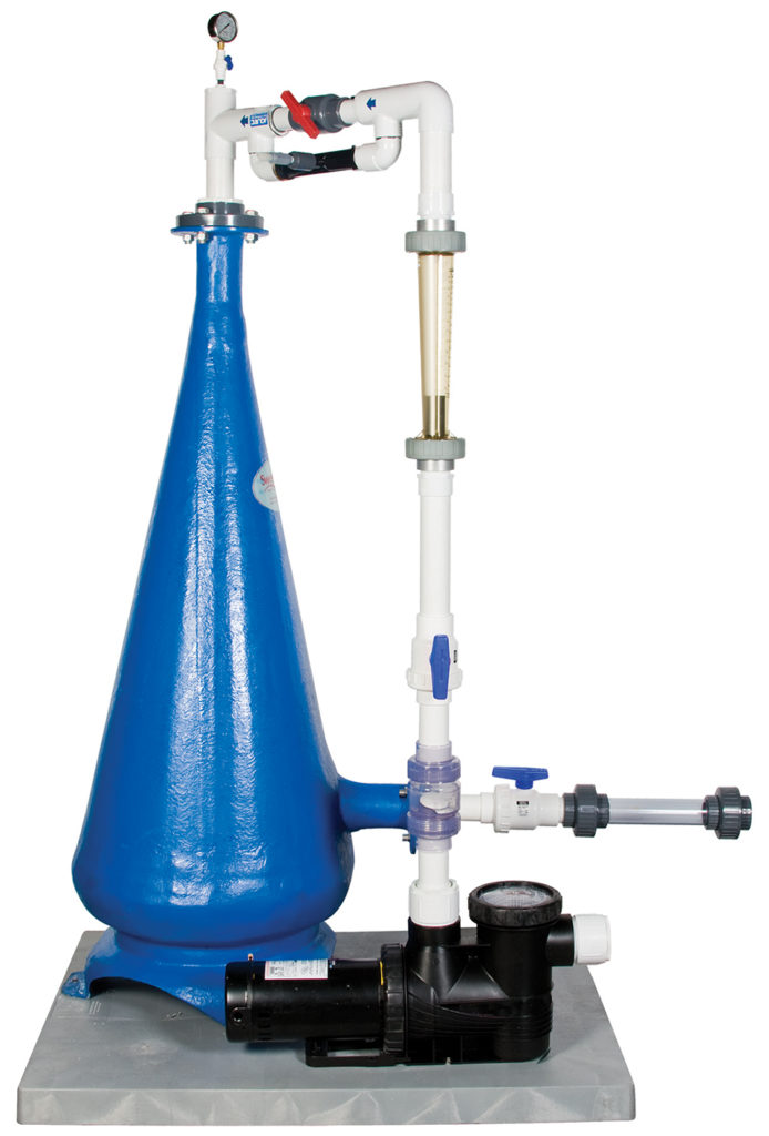
A look at unit processes in RAS systems
The ability to maintain adequate oxygen levels can be a limiting factor in carrying capacities for RAS. The amount of oxygen required is largely dictated by the feed rate and length of time waste solids remain within the systems.
Responsibility
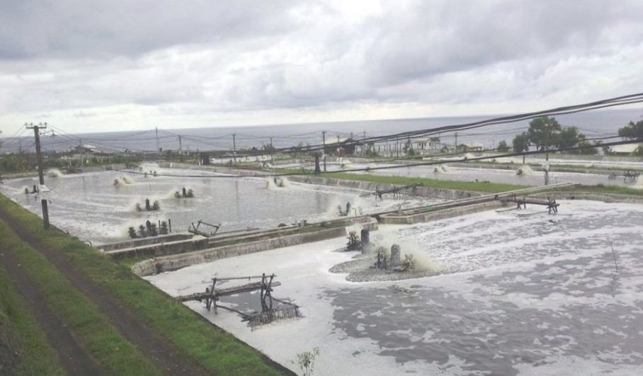
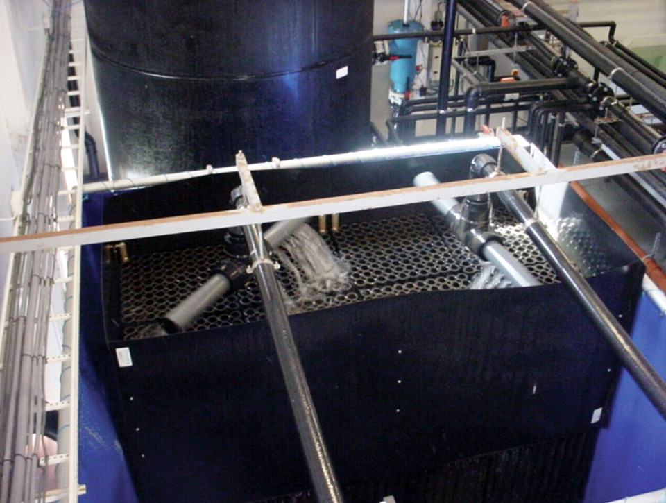
A look at various intensive shrimp farming systems in Asia
The impact of diseases led some Asian shrimp farming countries to develop biofloc and recirculation aquaculture system (RAS) production technologies. Treating incoming water for culture operations and wastewater treatment are biosecurity measures for disease prevention and control.
Innovation & Investment
A review of unit processes in RAS systems
Since un-ionized ammonia-nitrogen and nitrite-nitrogen are toxic to most finfish, controlling their concentrations in culture tanks is a primary objective in the design of recirculating aquaculture systems.